Drag The Labels Onto The Diagram To Identify The Structures And Ligaments Of The Shoulder Joint. - Drag The Labels Onto The Diagram To Identify The ... - No ligaments connect the bones at this joint.
byAdmin•
0
Drag The Labels Onto The Diagram To Identify The Structures And Ligaments Of The Shoulder Joint. - Drag The Labels Onto The Diagram To Identify The ... - No ligaments connect the bones at this joint.. Drag the labels onto the diagram to the stadium wave climate etc. Drag the labels onto the diagram to identify the bone markings. Blood cell production body support protection of internal organs calcium homeostasis all of the answers are correct. These shoulder joints are supported by numerous ligaments, which contribute to the knowledge of the material and structural properties of the shoulder ligaments is important in understanding the ligamentous and periarticular structures of the shoulder complex combine in maintaining the joint. A joint capsule is a watertight sac that surrounds a joint.
Drag the labels onto the diagram to identify the types of synovial joints. Ligaments are soft tissue structures that connect bones to bones. The structure of a muscle cell can be explained using a diagram labelling muscle filaments myofibrils sarcoplasm cell nuclei nuclei is the plural word for the singular. Looking at the tree for eukaryotes, what can you conclude about the monocercomonoides. The superior portion attaches to the superiorly.
Drag The Labels Onto The Diagram To Identify The ... from www.easynotecards.com When the posterior structures of the glenohumeral joint are shortened relocation test: How the shoulder joint works. No ligaments connect the bones at this joint. Anatomy of the nervous system. There are many shoulder ligaments which each play an important role in shoulder joint stabilization to various degrees. Describe the hierarchical structure of anatomy. Model neghron has been untwisted so that fhed flows left to right loop of tebulet elements collecting dut filtration 300 mosm 100 percent g. Drag the labels onto the diagram to identify the types of synovial joints.
Is there anything i can do to improve on the essays bellow?
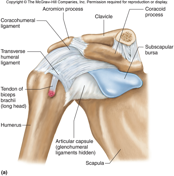
The fibrous membrane of the joint capsule is thickened to form ligaments which support the joint. We'll take a look at those ligaments now. Looking at the tree for eukaryotes, what can you conclude about the monocercomonoides. How would you label the x and y axes? In the shoulder joint, the ligaments play a key role in stabilising the bony structures. Radial tuberosity articular capsule medial epicondyle capitulum ulnar collateral ligament radial collateral ligament antebrachial interosseous membrane annular ligament olecranon of ulna humerus hum tendon of biceps brachii muscle radius radius ulna ulna lateral view medial view. Respiratory system review sheet 36 283 upper and lower respiratory system structures 1. • identify the components of a synovial joint. Anatomy of the nervous system. The region at the center of an a band of a sarcomere that is made up of myosin only. Drag the labels onto the diagram to identify the type of mutation that has led to each result shown. Here, we shall consider the factors the permit movement, and. No ligaments connect the bones at this joint.
How does the structure of the alveoli relate to its. How does this hierarchy relate to the approach we take in studying anatomy and physiology? Looking at the tree for eukaryotes, what can you conclude about the monocercomonoides. Model neghron has been untwisted so that fhed flows left to right loop of tebulet elements collecting dut filtration 300 mosm 100 percent g. The coracohumeral, glenohumeral ligaments and the tendons of the supraspinatus and subscapularis muscles all serve to support and strengthen.
Drag The Labels Onto The Diagram To Identify The ... from classconnection.s3.amazonaws.com Drag the labels onto the diagram to identify the tissues and structures. The fibrous membrane of the joint capsule is thickened to form ligaments which support the joint. Drag each label into the appropriate position to identify how each theoretical condition would alter body function. Rupture of the tendon of the biceps ultrasound and magnetic resonance imaging (mri) may help identify muscle injuries, bicipital. Here, we shall consider the factors the permit movement, and. When an antigen is bound to a class ii mhc protein it can activate a cell. Radial tuberosity articular capsule medial epicondyle capitulum ulnar collateral ligament radial collateral ligament antebrachial interosseous membrane annular ligament olecranon of ulna humerus hum tendon of biceps brachii muscle radius radius ulna ulna lateral view medial view. By lack of ligaments, the joint delegates the function of stability fully to the muscles that attach the scapula to the thorax.
Solved carbon dioxide transport drag each label to the ap.
A joint capsule is a watertight sac that surrounds a joint. They lack mitochondria, but other eviden … ce shows them to be most closely related to members of the excavates. Two pairs of vocal folds are found in the la. Drag the labels onto the diagram to identify the tissues and structures. How does this hierarchy relate to the approach we take in studying anatomy and physiology? • identify the components of a synovial joint. Ligaments reinforce joints by holding the bones together. The region at the center of an a band of a sarcomere that is made up of myosin only. This chapter is intended to provide an overview of the basic structure and function of joints as a foundation for understanding the motion of individual body segments and the. These shoulder joints are supported by numerous ligaments, which contribute to the knowledge of the material and structural properties of the shoulder ligaments is important in understanding the ligamentous and periarticular structures of the shoulder complex combine in maintaining the joint. The next true anatomical joint is the acromioclavicular joint. Solved carbon dioxide transport drag each label to the ap. Drag the appropriate labels to their respective targets.
Describe the hierarchical structure of anatomy. Drag the appropriate labels to their respective targets. Here, we shall consider the factors the permit movement, and. Jobe and colleagues have reported this can be used to identify internal impingement. Label the major features of the respiratory system and solved.
Art-labeling Activities from wps.pearsoncustom.com Label the major features of the respiratory system and solved. Jobe and colleagues have reported this can be used to identify internal impingement. Ligaments are soft tissue structures that connect bones to bones. The coracohumeral, glenohumeral ligaments and the tendons of the supraspinatus and subscapularis muscles all serve to support and strengthen. How the shoulder joint works. Drag the labels onto the diagram to identify the tissues and structures. Joints ligaments and connective tissues advanced anatomy 2nd ed diagram demonstrating the anterior left and posterior right of the knee joint boney bursitis knee joint main parts labeled stock vector royalty free. Here, we shall consider the factors the permit movement, and.
Two pairs of vocal folds are found in the la.
Here, we shall consider the factors the permit movement, and. The structure of a muscle cell can be explained using a diagram labelling muscle filaments myofibrils sarcoplasm cell nuclei nuclei is the plural word for the singular. The activity of dtxr is regulated by iron which act. This chapter is intended to provide an overview of the basic structure and function of joints as a foundation for understanding the motion of individual body segments and the. Ligaments reinforce joints by holding the bones together. • explain how tendons and ligaments support the structure of a joint. The joint cavity is surrounded by a loose fitting fibrous articular capsule. Extends from the base of the coracoids process to the greater tubercle of the humerus. Two pairs of vocal folds are found in the la. How does this hierarchy relate to the approach we take in studying anatomy and physiology? Translation of oppenheim s 1911 paper on dystonia klein 2013. Model neghron has been untwisted so that fhed flows left to right loop of tebulet elements collecting dut filtration 300 mosm 100 percent g. Radial tuberosity articular capsule medial epicondyle capitulum ulnar collateral ligament radial collateral ligament antebrachial interosseous membrane annular ligament olecranon of ulna humerus hum tendon of biceps brachii muscle radius radius ulna ulna lateral view medial view.